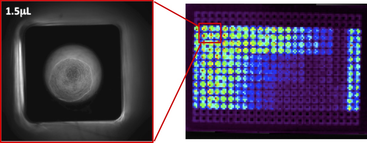
3-D models of epithelial tissues
The epithelium represents a major class of tissue architecture with unique functions. It serves as a regulator of mass transport, defence against pathogens, and source of mucosal extracellular matrix (ECM). Dysregulation of the normal turnover of epithelial cells has been associated with several chronic diseases, including fibrosis and cancer. The current gold standard for studying epithelium function in vitro is the transwell culture. However, the lack of physiologically relevant dimensional cues and mismatched material properties between cells and polymer membranes can lead to unnatural epithelium morphologies and functions. Our proposed solution is to use 3-D epithelial acini (i.e., well-organized and polarized spherical epithelial structures) as a long-term and physiologically relevant epithelium model. Currently we are using this platform to model epigenetic and metabolic changes in during epithelial dysplasia, which is characterized by uncontrolled cell growth and progressive tissue disorganization. We are also developing new tools to help monitor changes of epithelial function in real-time in response to microbial attacks. The resulting model will be broadly useful for studying epithelial tissues, including airway and digestive track.

Bio-printed human-microbe co-cultures
Human beings live alongside a vast variety of microorganisms in the environment, many of which reside within us and in direct contact with our mucosal epithelium (e.g oral, airways and gut). Recent research has highlighted the associations between microbes and human health, from metabolic diseases to cancers. However, little is known about the mechanisms of these interactions due to the lack of translatable in vivo and in vitro models. To date, few in vitro systems are able to directly co-culture human cells and microbes in a defined and repeatable fashion. Our group aims to address this problem by using advanced bio-printing techniques and specially formulated bio-ink to confine bacterial colonies in direct co-culture with engineered human tissues (see project 1). In an earlier study we have successfully cultivated bacteria biofilm on an epithelial cell monolayer using an aqueous two-phase system (ATPS). The goal here is to expand on this concept to create long-term, multiplexed bacteria colonies on a 3-D epithelium model to study human microbiome related diseases.

In vitro models of tumour microenvironment
Over the course of tumour development, its microenvironment becomes increasingly complex and develops significant materials gradients (e.g. glucose, oxygen etc.) due to diffusion limitation. In response, tumour cells develop a variety of coping mechanisms, including altered metabolic and signalling pathways. Some cells even take on specialized role as tumour initiating cells (TIC) that plays key roles in tumour survival and metastasis. However, current in vitro models cannot faithfully recreate the niche or maintain the metabolic profiles of TIC from explanted tumours. These challenges have severely limited the development of effective therapeutic strategies. In this proposed project, we aim to develop and validate a multi-layered tumour model by combining bioprinting technology, functionalized extracellular matrix and advanced biosensing techniques that delivers a realistic and programable tumour microenvironment. Our long term goal is to use such model to understand the dynamics of TIC and immune cells within the tumour microenvironment in order to better understand their signalling pathways and discover therapeutic targets.
Biomaterials design for dental pulp regeneration
Endodontic therapy, or commonly known as root canal therapy, is a standard procedure for treating infected pulp by replacing living pulp tissue with inert filling material. However, the loss of living pulp tissue prevents future growth and development of the dentinal wall, and this is particularly problematic for young patients as it leads to weak tooth and increase chances of fracture. Recently, regenerative endodontic therapy based on revascularization has gain traction as a promising option to restore dentin development. The technique relies on the formation of a blood clot in the root canal space to act as a provisional scaffold by mechanically inducing bleeding from the root. However, controlled formation of blood clot in situ is difficult, which may lead to unpredictable results. In collaboration with Dr. Isabel Mello (Dalhousie) we are developing novel biomaterials to guide the formation of blood clot and to assist the migration of stem cells of the apical papilla (SCAP) into the root canal. We are exploring the use of 3-D bioprinting technology to deliver cell migration cues and differentiation cues directly into the structure of biomaterials to direct SCAP function within the root canal. In addition, we are building in vitro models of the root canal to better understand the transport kinetics of antimicrobial and antiseptic agents so that we can better understand their roles in SCAP survival.

